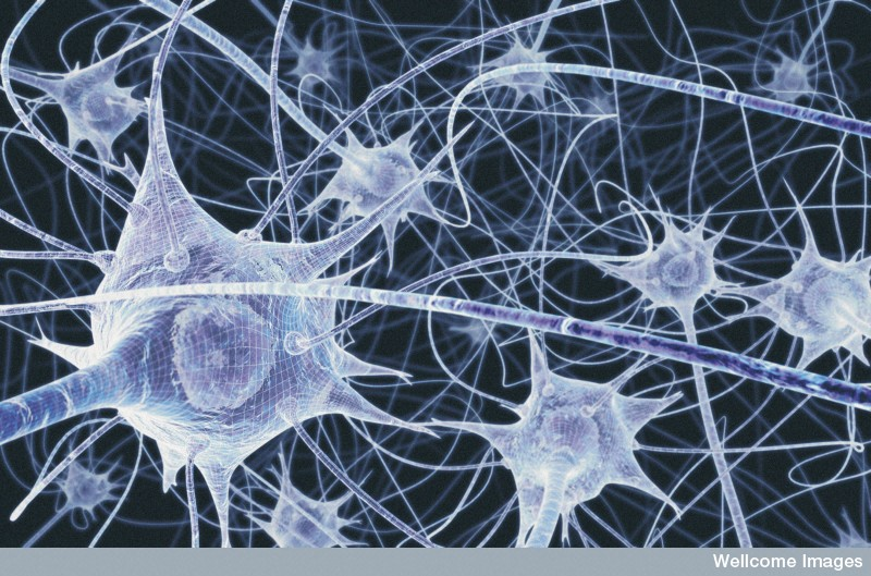
Transmission of Nerve ImpulsesThe human brain contains billions of neurons (nerve cells) arranged in a gigantic network (artist's impression, right). The neurons in this network communicate with each other by transmitting nerve impulses (signals) from one neuron to another. The process by which this is done is very complicated but surprisingly well understood. This project discusses how this transmission is carried out. Click on the picture to view the article in a separate tab. |
|

STAGE A: Synaptic Transmission
As already mentioned, synaptic transmission is the process that lets a nerve impulse pass from one neuron to the next across the synaptic gap (or just synapse). It is the first of the three steps in the transmission of a nerve impulse.
Synaptic transmission is a chemical transmission effected by neurotransmitters. A neurotransmitter is a molecule that moves across the synapse and, by binding (attaching) to a receptor on the
Transmission begins when an electrical impulse travelling along an axon reaches the
Ion channels
We have mentioned that a neuron membrane contains leakage channels which are open all the time. Membranes also have two other kinds of ion channels, as follows:
Ligand (or binding) ion channels: These channels are normally closed, but open when a neurotransmitter binds to the channel. The channel then allows ions to enter. See the diagram on the below.
It is these two kinds of channels that are involved in synaptic transmission. Note that the leakage channels are still present and still function (but are not shown in the diagram). However, as they are just 'leakage' channels. the number of ions that move through them is very small, whereas many ions move through the other two kinds of channels.
ligand ion channel
voltage- sensitive ion channel
Main steps in synaptic transmission by neurotransmitters
Chemical transmission takes place in the synapses between neurons, enabling nerve impulses to be transmitted from one neuron to the next. This process is called chemical transmission because the diffusion of chemical molecules from one neuron to the next is what enables an impulse to be reconstituted in that second neuron as an electrical impulse. Chemical transmission gives the brain the flexibility that is required for learning. The following are the main steps for synaptic transmission.
– 21 –

Step 1: Synthesis
The neurotransmitter is synthesised (made) in the terminal button of the axon from a precursor (i.e. a substance that is changed into another substance, in this case a neurotransmitter) using enzymes present in the axon.
(An enzyme is a substance that helps to speed up a chemical reaction without itself being changed.)
The neurotransmitters are then stored in
synaptic vesicles (a vesicle is a small
Step 2: Secretion
When a nerve impulse travelling down an |
|
|
|
|
|
|
|
|
|
|
|
|
|
|
|
|
|
Ca2+ |
||
|
|
|
|
|
|
|
|
|
|
|
|
|
|
|
|
|
ions |
|||
axon (also called an action potential as |
|
|
|
|
|
|
|
|
|
|
|
|
|
|
|
|
|
|
voltage- |
|
labelled in the diagram on the right) reaches |
|
|
|
|
|
|
|
|
|
|
|
|
|
|
|
|
|
|
sensitive |
|
the terminal button, it causes the voltage- |
|
|
|
|
|
|
|
|
|
|
|
|
|
|
|
|
|
|
Ca2+ ion |
|
ligand |
|
|
|
channel |
||||||||||||||||
sensitive Ca2+ channels to open, allowing |
|
|
|
|
|
|||||||||||||||
Na+ ion |
|
|
|
|
|
|||||||||||||||
many Ca2+ ions to enter the terminal |
channel |
|
|
|
|
|
||||||||||||||
|
|
|
|
|
|
|
|
|
|
|
|
|
|
|
|
|
|
- |
|
|
button. This causes the synaptic vesicles to |
|
|
|
|
|
|
|
|
|
|
|
|
- |
|
- |
- |
|
|||
move down and merge with the cell |
- |
|
- |
|
- |
|
- |
|
- |
|
- |
|
|
|
|
|
|
|
||
|
|
|
|
|
|
|
|
|
|
|
|
|||||||||
membrane, releasing neurotransmitters into |
|
|
|
|
|
|
|
|
|
|
|
|||||||||
|
|
|
|
|
|
|
|
|
|
|
|
|
|
|
|
|
|
|
|
|
the synaptic gap (the black dots represent the neurotransmitter molecules). The empty
vesicles then close up again and retreat inward, ready to be filled with neurotransmitters again.
Note: In this diagram, charges on the inner and outer layers of the membrane are shown. Remember that in a neuron at rest, the outer layer is positively charged (+) while the inner layer is negatively charged
Step 3: Binding
The neurotransmitters move across the synaptic gap and bind to receptors (ion channels) on the membrane of the post- synaptic neuron that are specific to that neurotransmitter. (In the diagram, the binding is shown by the small black dots – the neurotransmitters – sticking to the top of the receptors/ion channels.) This results in the ligand Na+ ion channels opening
– 22 –

and letting in sodium ions.
The small diagrams (on the right) show an enlarged view of what happens. The red dot is the neurotransmitter molecule binding to the Na+ ion channel. Then Na+ ions enter.
We have said earlier, that in the rest state, the inside of a neuron is negative (as shown in the diagram for Step 1). But after the Na+ ions pour in, there are now more positive charges than negative charges so the inside layer of the membrane now has an overall positive charge, as the lower small diagram shows. Note: On the outer layer of the membrane there is actually almost no change in the charge as there are billions of Na+ ions outside, so the movement of a few inside the dendrite makes practically no difference. However, the outside layer is now relatively negative compared to the inner layer of the membrane. So after the entry of the Na+ ions, there is a reversal of charge as the lower small diagram shows.
Step 4: Inactivation
The neurotransmitters unbind from the membrane and return to the synaptic gap where they are quickly inactivated to allow the receptors (ion channels) to be available for the next stimulus. Neurotransmitters can be inactivated by one or a combination of the following processes:
(1)They simply diffuse out of synaptic gap and spread into the space outside the neurons,
(2)They are broken down into bits by enzymes present in the synaptic gap,
(3)They are reabsorbed by the terminal button of the
(4)They are removed from the synaptic gap and destroyed by one kind of glial cells (known as astrocytes).
Later, in Stage B, we will see that the Na+ ions that have entered the
Extra: Electrical transmission across a synapse
Most synapses involve chemical transmitters as discussed above. But a few neurons involve electrical transmission across the synaptic gap. Some synapses are both electrical and chemical; at these, the electrical response occurs earlier than the chemical response.
An electrical synapse is often called a gap junction, in which the membranes of the two neurons are connected by channels that let the ions pass directly from one neuron to the other. Gap junctions allow for a more
– 23 –

rapid communication. But we will not be concerned with these.
Chemical synapses are slower than electrical ones but are also far more flexible (without going into what this means). This valuable flexibility is the foundation of all learning.
Strengthening the transmission: AMPA and NMDA receptors
The chemical transmission across the synapse can be made stronger, which results in a stronger electrical signal travelling down the rest of the
1.By increasing the number of receptors (ion channels) in the
which, for those interested, is short for
2.By using another type of receptor (called an NMDA receptor, which is short for
Both kinds of receptors are activated by glutamate neurotransmitter (red dots in above diagram) which binds to the receptors.
The AMPA receptor is only ligand gated, that is, it opens only when the neurotransmitter binds to it. The NMDA receptor however, is both voltage and ligand gated, that is, it depends on the inside of the membrane having a high enough positive charge (voltage) to eject the Mg2+ ion then binds with the neurotransmitter to allow ions to enter the neuron.
How the two kinds of receptors function depends on the strength of the stimulus/signal/nerve impulse (call it what you like!) arriving from the
1.Weak stimulation: That is, when only a few neurotransmitters cross the synaptic gap. This normally activates just the AMPA receptors. This is because the Mg2+ ions at the core of the NMDA channels block these channel thus preventing ions from entering through them. AMPA channels do not have this Mg2+ ion. The diagrams below show what happens.
The AMPA receptor is shown in blue, the NMDA receptor in pink (with the Mg2+ ion blocking its pore). The neurotransmitter crosses the synaptic gap and binds to the AMPA receptor, allowing Na+ ions to enter. While they may also bind to the NMDA receptor (as shown in the third diagram), the Mg2+ ion remains in place.
– 24 –

2.Strong stimulation: A strong stimulus occurs when there are lots of neurotransmitters or there is brief
The calcium ions (Ca2+) that enter the
[The Ca2+ ions also seem to play a part in the formation of our long- term memories though detailed mechanisms of how this happens are not yet known.]
– 25 –

Extension: Phosphorylation
Phosphorylation is the addition of phosphate ions
http://www.sumanasinc.com/webcontent/animations/content/ampa_and_nmda.html
Look at slides 5 – 8 in particular which elaborate on this and also provide some animations.
Video animation
How transmission across a synapse occurs. Here are two of many video animations available on the web:
https://www.youtube.com/watch?v=p5zFgT4aofA
https://www.youtube.com/watch?v=WhowH0kb7n0
Note the use of terms not refereed to in this text: electrical impulse = action potential, vesicle = container/sac that holds neurotransmitter molecules) See also diagram at right.
– 26 –